


Bio-Physics Week Lecture: Cryo-Electron Microscopy and Its Application
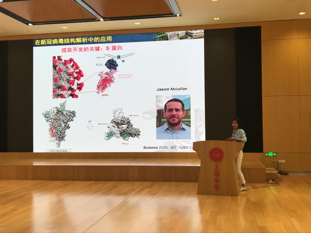
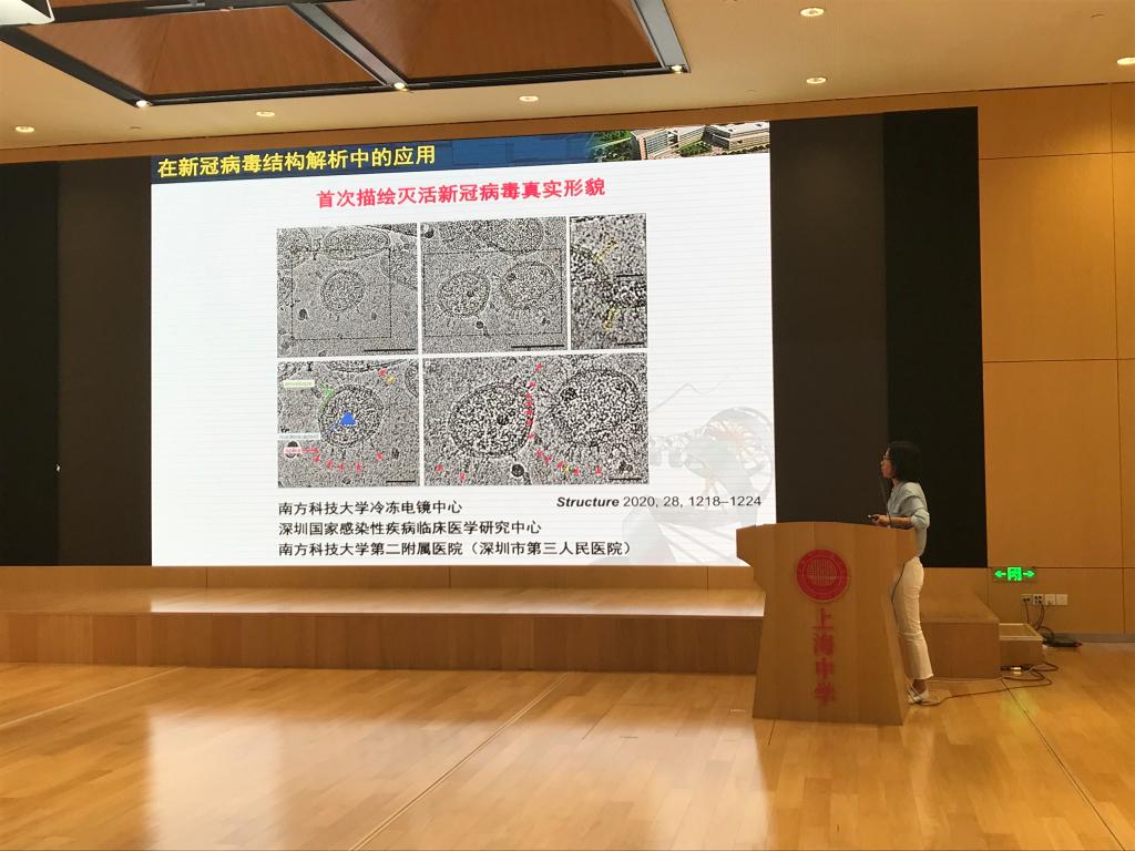
In the battle against the COVID-19 pandemic, one technique reveals the "look" of the virus to the world, which is of great importance in understanding the characteristics of the virus and developing vaccines. The technique, known as cryo-electron microscopy, allowed its developers to win the 2017 Nobel Prize in Chemistry.
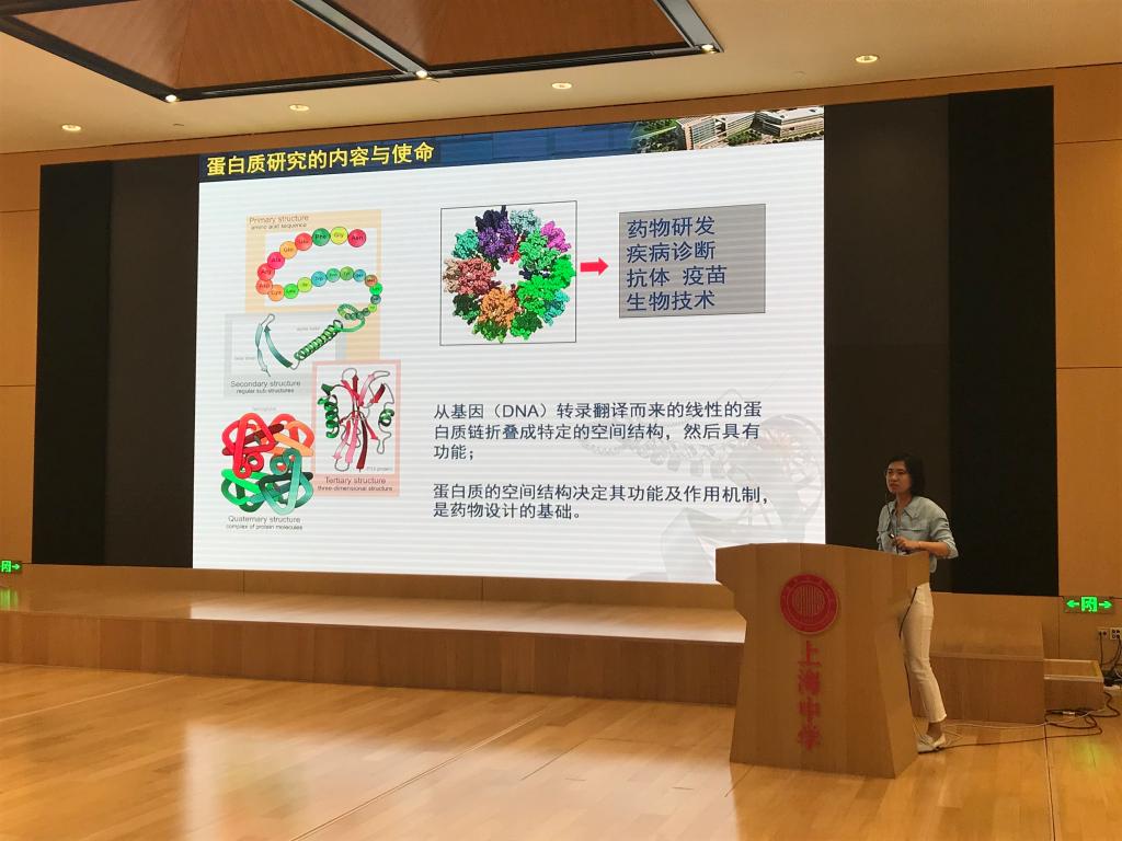
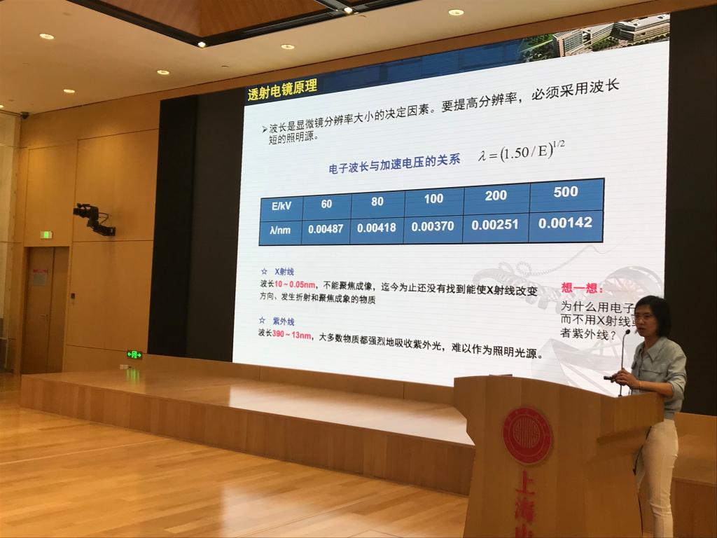
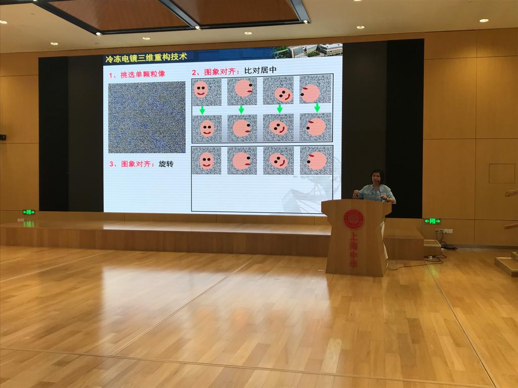
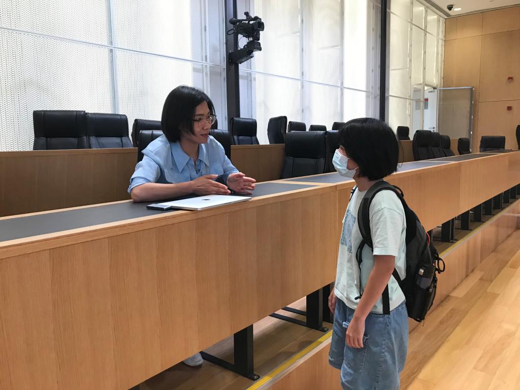
On June 3, Ms. Wang Fangfang from the National Center for Protein Science Shanghai gave a wonderful lecture to students and teachers at SHSID: Cryo-Electron Microscopy and Its Application in the Structural Analysis of the Novel Coronavirus 2019-nCoV. In the lecture, Ms. Wang introduced how cryo-electron microscopes work and their application in a vivid and easy-to-understand way. The lecture started with the importance of analyzing the 3D structure of proteins, then compared three methods of studying protein structures, and introduced the background under which cryo-electron microscopy was invented and developed. In order to let students better understand the working principle of cryo-electron microscopes, Ms. Wang started with transmission-electron microscopes, and used light microscopes, which students were familiar with, as a comparison. Through Ms. Wang’s detailed instruction on how to prepare samples, students seemed to be on the scene and saw how a virus sample added to the copper grid was processed step by step into a sample ready to be observed. Questions asked by Ms. Wang from time to time, such as what vitreous ice was and why liquid ethane was used instead of liquid nitrogen for freezing samples, made the lecture more fascinating and also allowed students to better understand the content. The part that had interested students the most might have been how to construct a 3D structure from 2D images observed by a cryo-electron microscope. In this part, Ms. Wang replaced images of viruses with 2D cross-sections of familiar 3D objects such as houses, and vividly demonstrated this process. The products of cryo-electron microscopy, such as the high-definition 3D animation of the COVID-19 virus, also instantly attracted the attention of students. After the lecture, students asked questions actively and stayed for further discussion with Ms. Wang.
The cryo-electron microscopy developed by physicists has promoted the rapid development of structural biology, making students see the importance of interdisciplinary thinking. At the same time, Ms. Wang’s in-depth yet clear explanations showed students the basic scientific principles behind complex techniques as well as the application of what they have learned.
(Written by Chengbo Zhou Pictures by Chengbo Zhou Reviewed by Qian Zuo)

- No.989, Baise Road, Shanghai, China, 200231
- No.38, Huoxiang Road(Pudong Campus), Shanghai, China, 201203
- © 2018 Shanghai High School International Division